Screenshots
Main database
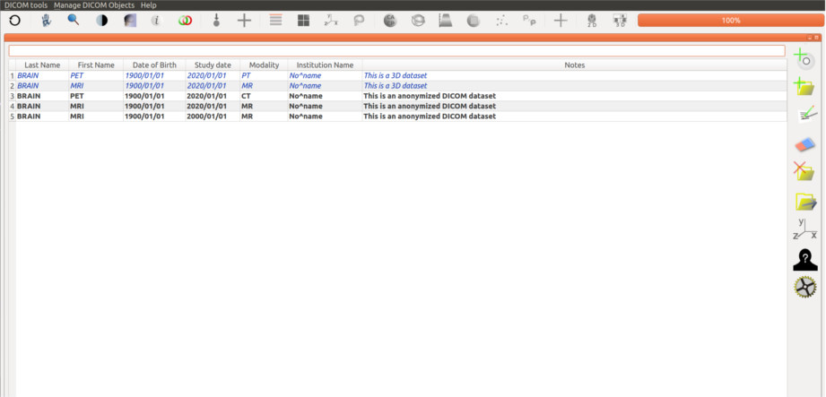
The main database gives the access to the DICOM datasets (double-click on image to enlarge). It also provides:
- an anonymisation algorithm,
- an extraction tool to transfert DICOM data to an external USB disk,
- a free space for recording personal notes.
2D DICOM image reader
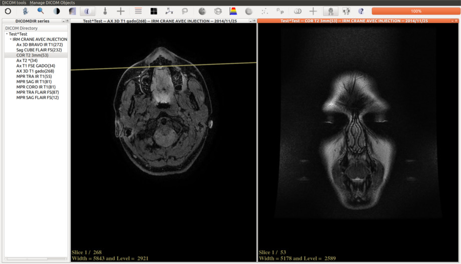
- The number of display windows can be set from 1 to 9.
- Use drag and drop to select a DICOM serie.
- The yellow line shows the projection of the intersection between the selected image and the other images.
Oblique reslicing
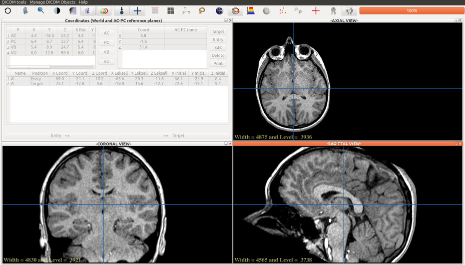
During navigation in a DICOM 3D dataset, a sustained right mouse click will initialise a rigid rotation according to the angle defined by the course of the mouse from its initial position to its final course. The center of the rotation is the last location of the user inside the volume.
Multi modal registration
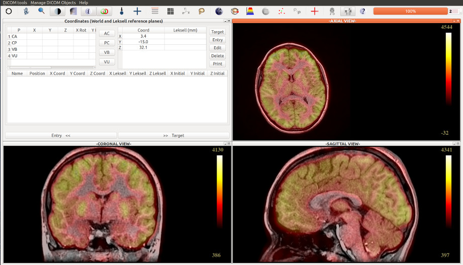
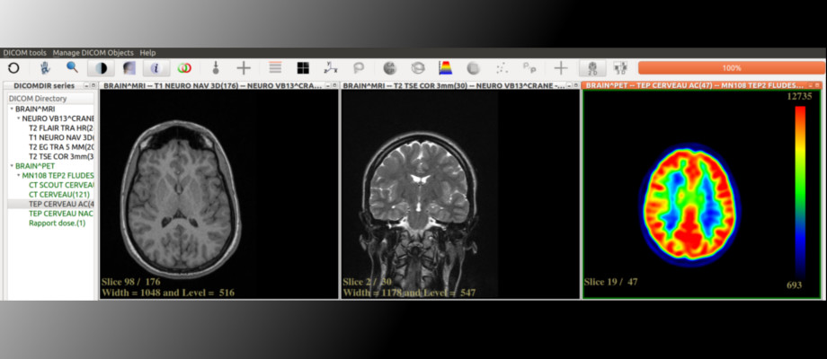
Multi modal registration is enabled:
- IRM/TDM.
- IRM/PET scan,
Color tables are proposed. Red-yellow table in the example above (click to enlarge).
2D Brain depth electrodes visualization
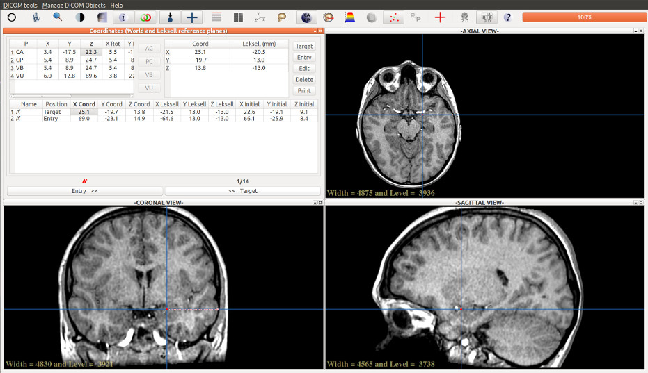
- The user select the entry and the target points.
- Navigation through the depth electrodes is enabled from the entry to the target points using the navigation buttons.
- Left electrodes are colored in red, right electordes are colored in green.
- The data are stored memory for further access.
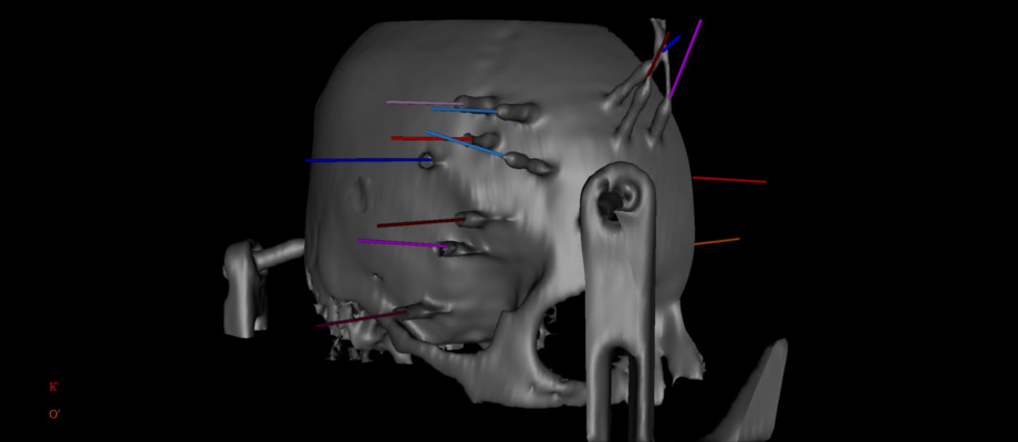
3D Brain depth electrodes visualization
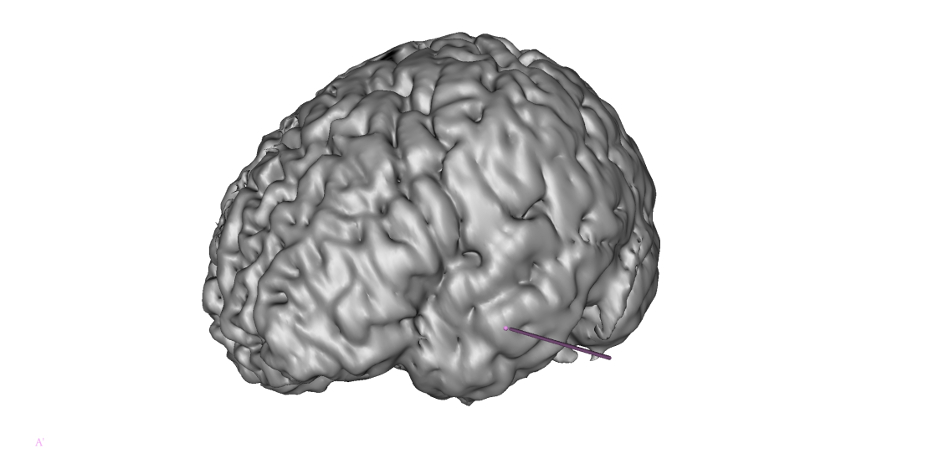
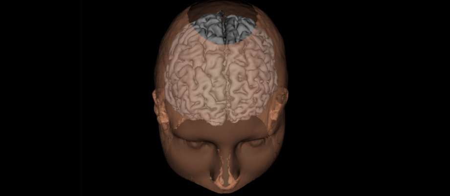
3D depthelectrodes visualization is provided using a brain extraction tool and a density algorithm.
In the example above, visualization of the brain left gyri with a brain depth elecrode targeting the left amygdala with an entry point in the left anterior part of T2 temporal gyrus.
